What does PEMF Produce at the Cellular Level?
PEMF Increases Cellular Membrane Permeability
As early as 1940, it was suggested that magnetic fields affect the TMP and the flow of ions in and out of the cells and might therefore influence cellular membrane permeability. It has since been established that Pulsed Electromagnetic Fields (PEMF) can influence ATP (Adenosine Tri-phosphate) production; increase the supply of oxygen and nutrients; improve the removal of waste and help rebalance the distribution of ions across the cell membrane.
Healthy cells in tissue have a voltage difference between the inner and outer membrane referred to as the membrane resting potential that ranges from -70 to -90 mV. This causes a steady flow of ions through its voltage-dependent ion channels. In a damaged cell, the potential is raised, and an increased sodium inflow occurs. As a result, interstitial fluid is attracted to the inner cellular space, resulting in swelling and edema. The application of PEMF to damaged cells accelerates the reestablishment of normal potentials (Sanseverino, 1999) increasing the rate of healing and reducing swelling.
HEALTHY CELL

UNHEALTHY CELL
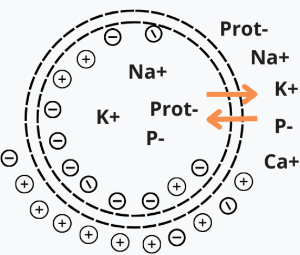
If a cell has a resting potential of -70mV and the membrane potential rises to -50mV, then the cell has been depolarized. Depolarization is often caused by an influx of cations, e.g. Na+ through Na+ channels, or Ca2+ through Ca2+ channels. On the other hand, the efflux of K+ through K+ channels inhibits depolarization, as does an influx of Cl– (an anion) through Cl– channels. If a cell has K+ or Cl– currents at rest, then inhibition of those currents will also result in depolarization.
If a cell has a resting potential of -70mV and the membrane potential rises to -50mV, then the cell has been depolarized. Depolarization is often caused by an influx of cations, e.g. Na+ through Na+ channels, or Ca2+ through Ca2+ channels. On the other hand, the efflux of K+ through K+ channels inhibits depolarization, as does an influx of Cl– (an anion) through Cl– channels. If a cell has K+ or Cl– currents at rest, then inhibition of those currents will also result in depolarization.
As the Pulsed Electromagnetic Field temporarily hyperpolarizes and depolarizes the membrane, the ion channels open and close allowing a more efficient ion exchange, as with the sodium-potassium (Na+, K+) pump, thus increasing cellular oxygenation and nutrition as sodium export stimulates several secondary active transporters.
PEMF Increases Cell Metabolism
In a study on Chronic Fatigue Syndrome and Electromedicine, Thomas Valone, Ph.D., showed that damaged or diseased cells present an abnormally low TMP, about 80% lower than healthy cells. This signifies a greatly reduced metabolism and impairment of the electrogenic Na+/ K+ pump activity associated with reduced ATP (Adenosine Tri-Phosphate = energy) production.
The sodium-potassium pump uses energy derived from ATP to exchange sodium for potassium ions across the membrane. An impaired Na+/ K+ pump results in edema (cellular water accumulation) and a tendency toward fermentation, a condition known to be favorable toward cancerous activity.
French researcher Louis C. Kervran demonstrated that Sodium plus Oxygen plus Energy (ex: PEMF) nuclearly transmutes into Potassium as follows: 11 Na23 + 8 O16 + energy = 19 K39 This nuclear process is accomplished with low heat, which is the most important and commonly occurring phenomenon of Nuclear Fusion in Biology.
PEMF provides the energy to produce this natural phenomenon in the body. This is why PEMF is an excellent treatment for high blood pressure.
As a result of a better utilization of oxygen in the cells, they (the cells) increase the production of their energy (ATP). The organism becomes more stable and efficient; toxins and waste products are broken down and eliminated more quickly. The body's natural regulatory mechanisms are strengthened, and healing processes are accelerated.
PEMF is a strong antioxidant. First, we take a look at what it is a free radical. It is a type of unstable molecule that is produced during normal cellular metabolism (chemical changes that occur in a cell). Free radicals can accumulate in cells and damage other molecules, such as DNA, lipids, and proteins. This damage can increase the risk of cancer and other diseases.
Free radical proliferation is related to pathological changes that cause cell malfunction or mutation.
According to studies, free radicals also "deplete cellular energy" by interfering with mitochondrial function.
Cellular energy generation in mitochondria is both a key source and a key target of oxidative stress in cells.
Free radicals set off chain reactions when electrons are stripped from molecules, creating another free radical.
Antioxidants such as vitamin A, vitamin E, selenium, and coenzyme Q10 supply electrons to neutralize free radicals, without breaking down, and are often prescribed to provide relief and counter the ravages of free radicals.
However, PEMF therapy can also inhibit and eliminate free radicals by supplying abundant electrons to neutralize them.
You can read more about it in (Thomas Valone, Ph.D. in Bioelectromagnetic, 2003).
On the biophysical level, as PEMF therapy increases the circulation of electrons across the membrane, a parallel phenomenon seems to occur: the acceleration of ATP synthesis and of other aspects of the cell's biochemical anabolism. As electrons are drawn to the inner membrane, they increase the ionic charge inside the cell and, thus, the TMP. In 1976, Nobel Prize winner Dr. Albert Szent-Gyorgi established that structured proteins behave like diodes or rectifiers. A diode passes electricity in only one direction.
He proposed that cell membranes can rectify an induced voltage and this rectifying property of cell membranes can cause changes in the ion concentration of the inner and outer surfaces of the cell membrane in such a way as to increase the TMP and effectively stimulate the activity of the Na+/ K+ pump. Cell health is directly affected by the health of the Na+/ K+ pump, which is directly proportional to the TMP. Based on these biophysical principles, PEMF Therapy with sufficient intensity will stimulate the TMP, normal cell metabolism, the sodium pump, ATP production and healing.
“TMP is proportional to the activity of this pump and thus to the rate of healing.” Furthermore, “increases in the TMP have also been found to increase the uptake of amino acids.” (Dr. Albert Szent-Gyorgi). This is important, as increasing the supply of nutrients is also an effective aid to cell repair. This is particularly true in trauma where circulation has been impaired by crushed or severed blood vessels, or by the inflammation and swelling that compress capillaries, blocking the flow to both the injured and uninjured cells.
PEMF Increases Cellular Membrane Flexibility and Elasticity
A study entitled ―Modulation of collagen production in cultured fibroblasts by a low-frequency Pulsed Electromagnetic Field, by Murray et al (Biochim Biophys Acta) shows that the total protein synthesis was increased in confluent cells treated with a Pulsed Electromagnetic Field for the last 24 hours of culture, as well as in cells treated for a total of 6 days. However, in 6-day-treated cultures, collagen accumulation was specifically enhanced as compared to total protein, whereas after short-term exposure, collagen production was increased only to the same extent as total protein. These results indicate that a Pulsed Electromagnetic Field can specifically increase collagen production, the major differentiated function of fibroblasts, possibly by altering cyclic-AMP metabolism.
PEMF therapy successfully increases membrane flexibility by increasing the synthesis of collagen, a crucial protein that supports membrane elasticity, within the fibroblasts. In doing so, PEMF therapy increases tissue and muscle flexibility and, in doing so, increases range of motion.
PEMF Stimulates Cellular Communication and Replication
DNA synthesis is linked to low Pulsed-intensity Electromagnetic Fields (Liboff et al, 1984; Rosch et al, 2004). Proteins are conductors of electricity. When exposed to PEMF, proteins are subject to electrophoresis. The Ribonucleic Acid messengers (RNAm) that are synthesized from a Deoxyribonucleic Acid (DNA) template during transcription mediate the transfer of genetic information from the cell nucleus to ribosomes in the cytoplasm and serve as a template for protein synthesis.
Since RNA mechanically influences the DNA and encoded proteins influence RNA, the flow of information to and from genes may be linked to changing Pulsed Electromagnetic Fields (Einstein, 1977; Goodman et al, 1983). Since Pulsed Electromagnetic Fields interact with changing electrical charges (recent studies by Dandliker et al, 1997) show that DNA conducts electrons along the stacked bases within the DNA double helix, Pulsed Electromagnetic Fields may initiate transcription of the precursor mRNA by accelerating electrons moving within the DNA helix (McLean et al, 2003).
PEMF Increases Cellular Genesis (Cellular Growth and Repair)
The many intra and inter-cellular processes and activity stimulated by PEMF therapy lead to faster cellular and tissue regeneration. This fact is shown by the results of many studies on a variety of tissues, including bones, spine, cartilage, intestines, blood vessels, nerves, brain, and muscles.
In December 2004, the Swiss Medical Tribune stated that PEMF therapy provided: “improvement of blood circulation, relief from pain, improvement of bone healing and the stimulation of nerve cells. Not only is PEMF therapy effective in disease conditions: but it is also an excellent means of preventing stress and assisting regeneration and recovery after sports exertion. Through metabolic activation and blood circulation, more nutrients and oxygen are available to muscle cells, less damage is experienced, and efficiency is improved.
Other medical studies on Spine fusion for discogenic low back pain showed how PEMF relieves the pain and improves healing.
Marks RA. (Richardson Orthopaedic Surgery, TX, USA) randomly selected 61 patients who underwent lumbar fusion surgeries for discogenic low back pain between 1987 and 1994 and had failed to respond to preoperative conservative treatments. Average follow-up time was 15.6 months postoperatively. Fusion succeeded in 97.6% of the 42 patients who received PEMFtherapy for only 52.6% of the 19 patients who did not receive PEMF therapy stimulation of any kind.
A similar study by Richard A. Silver, M.D. (Tucson Orthopaedic & Fracture Surgery Associates, Ltd., Tucson, AZ, USA) with 85 patients who had undergone surgery of posterior lumbar interbody fusion (PLIF) and had risk factors associated with a poor prognosis for healing, including smoking, prior back surgery, multiple spinal levels fused, diabetes mellitus, and obesity, radiographic examination and clinical evidence indicated that all but two patients achieved successful fusion. Of the 83 patients with successful spinal fusion, 29 (34.9%) were assessed as "excellent," 45 (54.2%) as "good," 3 (3.6%) as "fair", and 6 (7.2%) as "poor". Adjunctive treatment with PEMF appeared effective in promoting spinal fusion following PLIF procedures across all patient subgroups.
PEMF on bone and cartilage in a study entitled: ―Modification of biological behavior of cells by Pulsed Electromagnetic Fields, 20 subjects of ages between 57 and 75 years with decreased bone mineral density, as defined by a bone densitometer, were treated with PEMF therapy during 12 weeks by Ben Philipson, (University of Hawaii School of Medicine, HI, USA). After 6 weeks, the bone density rose in those patients with an average of 5.6%. Properly applied Pulsed Electromagnetic Fields, if scaled for whole-body use, have clear clinical benefits in the treatment of bone diseases and related pain, often caused by micro-fractures in vertebrae. In addition, joint pain caused by worn-out cartilage layers can be treated successfully, through electromagnetic stimulation. PEMF application promotes bone union by electric current induction, which changes the permeability of cell membrane allowing more ions across, affects the activity of intracellular cyclic adenosine monophosphate (cAMP) and cyclic guanosine monophosphate (cGMP), and accelerates osteoblast differentiation by activation of p38 phosphorylation. PEMF stimulation also increases the partial oxygen pressure and calcium transport.
Repair and growth of cartilage is thus stimulated, preventing the grinding of the bones.
Overview of signal transduction pathways
A recent study on postoperative recovery led to the conclusion that PEMF therapy significantly reduced postoperative pain and narcotic use in the immediate postoperative period. Through a PEMF effect on nitric oxide signaling, which could impact the speed and quality of wound repair (Rohde et al, June 2009, Plastic & Reconstructive Surgery, Columbia, NY).
Nitric oxide is one of the few gaseous signaling molecules and a key vertebrate biological messenger that plays a role in a variety of biological processes. Recent studies show how PEMF therapy stimulates and rebalances many of these processes. There are several mechanisms by which nitric oxide has been demonstrated to affect the biology of living cells including oxidation of iron-containing proteins such as ribonucleotide reductase and aconitase, activation of the soluble guanylate cyclase, a single transmembrane protein, ADP ribosylation of proteins, a process of protein modification involved in cell signaling and the control of many cell processes including DNA repair, protein sulfhydryl group nitrosylation, another protein modification process, and iron regulatory factor activation. Having a lifetime of a few seconds, nitric oxide is highly reactive and diffuses freely across cell membranes.
These attributes make nitric oxide an ideal transient paracrine (between adjacent cells) and autocrine (within a single cell) signaling molecule. PEMF therapy is proven to effectively stimulate paracrine and autocrine communication.
Nitric oxide is also generated by phagocytes (monocytes, macrophages, and neutrophils) and, as such, is part of the human immune response.
Nitric oxide has been demonstrated to activate NF-κB in peripheral blood mononuclear cells, an important protein complex that controls the transcription of DNA and a transcription factor in iNOS gene expression in response to inflammation. It plays a key role in regulating the immune response to infection and is implicated in processes of synaptic plasticity and memory. The endothelium (inner lining) of blood vessels uses nitric oxide to signal the surrounding smooth muscle to relax, thus resulting in vasodilatation and increasing blood flow. As blood flow increases, so does the oxygen intake. PEMF therapy has proven to effectively increase blood flow and provide muscle relaxation, with better oxygenation of the muscle tissue.
Benefit of PEMF on Chronic Pain
In the chronic pain state, pain signal generation can occur in the central nervous system without peripheral noxious stimulation. In pain management, modulation of the pain signal transmission is a far better choice than neural destruction, and this can be achieved with PEMF.
Scientific evidence shows that acute persistent pain eventually sensitizes wide dynamic neurons in the dorsal horn of the spinal cord, the wind-up phenomenon, constituting the basis of developing chronic pain syndromes (Kristensen, 1992). Persistent and excessive pain has no biological good or necessary function. It is harmful to our well-being. Therefore, pain needs to be treated as early and as completely as possible and not be left alone (Adams et al 1997). The primary symptom in most patients with disorders affecting the soft tissue is pain. In many patients, daily activities are limited as pain causes a restriction of the range of movements. Causes of soft tissue pain can be depicted as musculoskeletal, neurologic, vascular, and referred to as visceral-somatic or articular (Cailliet, 1991). Early reports of applying electrical current to treat pain date back to before 1800 (Ersek, 1981). PEMF therapy has successfully been used for the control of pain associated with rotator cuff tendinitis, multiple sclerosis, carpal tunnel syndrome, and periarthritis (Battisti et al, 1998; Lecaire et al, 1991). An improvement was observed in 93% of patients suffering from carpal tunnel pain and in 83% in cases of rotator cuff tendinitis.
PEMF therapy was also used for the treatment of migraine, chronic pelvic pain, neck pain, and whiplash injuries (Rosch et al, 2004).
In a March 2003 publication on Pain Management with PEMF Treatment, Dr. William Pawluk explains, that Pulsed Electromagnetic Fields affect pain perception in many different ways. These actions are both direct and indirect.
The direct effects of Pulsed Electromagnetic Fields are on neuron firing, calcium ion movement, membrane potentials, endorphin levels, nitric oxide, dopamine levels, acupuncture actions, and nerve regeneration. Indirect benefits of Pulsed Electromagnetic Fields on physiologic function are circulation, muscle, edema, tissue oxygen, inflammation, healing, prostaglandins, cellular metabolism, and cell energy levels.
Short-term effects are thought to be due to a decrease in cortisol and noradrenaline, and an increase in serotonin, endorphins, and enkephalins. Longer-term effects may be due to CNS and/or peripheral nervous system biochemical and neuronal effects in which correction of pain messages occur, and the pain is not just masked as in the case of medication.
PEMF Blocks Pain
PEMF therapy has been shown to be effective at reducing pain both in the short-term and in the long term. The ways by which PEMF therapy relieves pain include pain blocking, decreased inflammation, increased cellular flexibility, increased blood and fluids circulation, and increased tissue oxygenation.
The trans-membrane potential, (TMP) is the voltage difference (or electrical potential difference) between the interior and exterior of a cell. An electrochemical gradient results from a spatial variation of both an electrical potential and a chemical concentration across a membrane. Both components are often due to ion gradients, particularly proton gradients, and the result is a type of potential energy available for cellular metabolism.
This can be calculated as a thermodynamic measure, an electrochemical potential that combines the concepts of energy stored in the form of chemical potential, which accounts for an ion's concentration gradient across a cellular membrane, and electrostatics, which accounts for an ion's tendency to move relative to the TMP.
Differences in concentration of ions on opposite sides of a cellular membrane produce the TMP. The largest contributions usually come from sodium (Na+) and chloride (Cl–) ions which have high concentrations in the extracellular region, and potassium (K+) ions, which along with large protein anions have high concentrations in the intracellular region. Opening or closing of ion channels for ion transport (Na+, Ca2+, K+, Cl-) in and out of cells at one point in the membrane produces a local change in the TMP, which causes an electric current to flow rapidly to other points in the membrane that occurs with the movement of electrons.
In a lecture on Pain Reduction, Dr. D. Laycock, Ph.D. Med. Eng. MBES, MIPEM, B.Ed., inspired by the works of Adams et al (1997) explain how PEMF therapy affects pain transmission at the levels of the neurons.
It is necessary to understand the mechanism of pain transmission to understand how pain blocking can take place with PEMF therapy. Pain is transmitted along the nerve cells by an electric signal. This signal encounters synaptic gaps at intervals. The pain signals are transmitted along nerve cells to pre-synaptic terminals. At these terminals, channels in the cell alter due to a movement of ions. The TMP changes, causing the release of a chemical transmitter from a synaptic vesicle contained within the membrane. The pain signal is chemically transferred across the synaptic gap to chemical receptors on the post-synaptic nerve cell. This all happens in about 1/2000th of a second, as the synaptic gap is only 20 to 50 nm (nanometers) wide. As the pain signal, in chemical form, approaches the post - synaptic cell, the membrane changes and the signal is transferred. During quiescent times, cells possess a small charge of about –70mV between the inner and outer membranes. When a pain signal arrives, it temporarily depolarizes the nociceptive cell and raises the cell TMP to +30mV. This increase is sufficient to open channels in the cell membrane allowing the exchange of the sodium (Na+) and potassium (K+) ions.
When an action potential begins, the channels that allow crossing of the Na+ ions open up. When the Na+ channels open, depolarization occurs, the Na+ rushes in because of both the greater concentration of Na+ on the outside and the more positive voltage on the outside of the axon. The flow of positively charged ions into the axon leads the axon to become positively charged relative to the outside. With each positively charged Na+ ion that enters the axon, another positive charge is inside and one fewer negative charge is outside the axon. Thus, together the inside grows increasingly more positive and the relative concentration of Na+ inside the axon relative to outside the axon grows greater. This initial phase of the action potential is called the depolarization phase. Now as the depolarization phase progresses, the status of the two physical forces that have been discussed changes. At the end of the depolarization phase, the voltage of the inside of the axon relative to the outside is positive and the relat ive concentration of Na+ ions inside the axon is greater than at the beginning of the action potential. The inside of the axon becomes sufficiently positive, about +30 mV as an average value, the Na+ channels close. This closing of the Na+ channels will greatly limit the ability of Na+ ions to enter the axon. In addition to the Na+ channels closing, the potassium (K+) channels open. Now K+ ions are free to cross the channels and now leave the axon due both to the greater concentration of K+ on the inside and the reversed voltage levels. The action potential is therefore not the movement of voltage or ions but the flow of these ion channels opening and closing moving down the axon.
This movement of the ion channels explains why the action potential is transferred slowly relative to the normal flow of electricity. The normal flow electricity is the flow of electrons in an electrical field and the electrons travel at the speed of light while the movement of these ion channels opening and closing is considerably slower. These are mechanical movements that cannot move as fast as the speed of light.
The exchange of the sodium (NA+) and potassium (K+) ions then triggers exocytosis of neurotransmitters via synaptic vesicles. These neurotransmitters diffuse into the synaptic gap. Once this process has occurred, the cell depolarizes back to its previous level of –70mV.
Research by Warnke established that the application of PEMF therapy has an effect on the quiescent potential of the neuronal synaptic membrane (Warnke, 1983; Warnke et al 1997). “It suggested that the effect is to lower the potential to a hyperpolarized level of –90mV. When a pain signal is received, the TMP has to be raised again in order to fire an action potential via neurotransmitters but it only achieves to raise the cell TMP to an approximate +10mV. This potential is well below the threshold of +30mV necessary to release the relevant neurotransmitters into the synaptic cleft and the pain signal is effectively blocked”.
By causing a hyperpolarized state at the neuronal membrane, PEMF therapy effectively blocks pain as it prevents the threshold necessary to transmit the pain signal to be reached.
Similarly, PEMF therapy effectively increases the TMP of damaged cells thus allowing them to recover their functions, heal and improve their metabolism.
It has been demonstrated that PEMF therapy effectively reduces pain in the short and long-term.
PEMF Decreases Inflammation
Several factors may contribute to inflammation including injury, tissue damage, a poor localized circulation with the formation of edema. Inflammation causes pain.
Swelling and bruising is an inflammation and discoloration of soft tissue caused by an impact injury or trauma. It can also result from surgery. Tissue cells are inherently like tiny electrically charged machines. When a cell is traumatized, the cell’s electrical charge is diminished; this causes normal cell functions and operations to shut down. Cells that are scarred or fibrotic with adhesions have a TMP charge of approximately -15 mV, degenerative or immune compromised cells average -30 mV, both low TMPs.
With the raised TMP, the body releases chemical signals that cause inflammation, swelling and bruising resulting in pain and inhibiting the cell communication pathways necessary for healing to begin. Numerous clinical studies have demonstrated that PEMF therapy has been successful in reducing inflammation. PEMF therapy treats the cellular source of swelling by recharging the cells with a mild electromagnetic current. This stops the release of pain and inflammatory mediators, reduces inflammatory fluids and allows an increase in blood flow, therefore increased oxygen intake, to help the cells heal faster with less swelling, pain and bruising.
In a study on the effect of wound healing Pulsed Electromagnetic Field on inflammatory cytokine gene expression in rats, Jasti et al (2001) states: ―Inflammation is characterized by massive infiltration of T lymphocytes, neutrophils and macrophages into the damaged tissue. These inflammatory cells produce a variety of cytokines, which are the cellular regulators of inflammation.
In a study on Low Frequency PEMF—a viable alternative therapy for arthritis published in 2009, Ganesan et al (Department of Biotechnology, Chennai, India) declare: ―PEMF for arthritis cure has conclusively shown that PEMF not only alleviates the pain in the arthritis condition but it also affords chondroprotection, exerts anti-inflammatory action and helps in bone remodeling, and this could be developed as a viable alternative for arthritis therapy.
Damaged cells are also energy deficient; thus they have low oxygen levels, high in sodium levels, and have a faltered electrochemical gradient. By inducing a mild electrical current into damaged cells, PEMF therapy slows or stops the release of pain and inflammatory mediators, increases blood flow, and re-establishes normal cell interaction. PEMF stimulates and restores the electrochemical gradient, the cell starts pumping sodium out, potassium enters the cell, the swelling resolves, oxygen starts flowing back in, and pain improves. Due to the density of the cell tissue, the Pulsed Electromagnetic Fields have to be strong enough (amount of energy of PEMF) to be able to restore the healthy TMP to its optimal -70 mV.
Several factors influence tissue inflammation and the processes by which PEMF therapy operates to reduce inflammation; include complex mechanical, chemical, electrical processes, along with increased circulation, oxygenation and cellular activity. With reduced inflammation, pain decreases and faster tissue healing occurs.
PEMF Increases Blood and Lymphatic Circulation
The arterial and venous blood vessels are intimately associated with the lymphatic system in the surrounding tissues of the body. As the blood and lymphatic vessels bring oxygen and nutrients to the cells and remove their waste products, they are nourishing and detoxifying the cells, tissues and body. PEMF therapy mechanically stimulates blood vessels and blood flow, the blood vessels pump blood and oxygen into the cells. Simultaneously, PEMF therapy mechanically stimulates the lymphatic vessels and waste products are hauled away from the cells more efficiently. PEMF therapy supports immune health by mechanically stimulating lymphatic drainage and blood flow.
In June 2004, The Faseb Journal states: “PEMF therapy has been shown to be clinically beneficial in repairing bones and other tissues, but the mechanism in action is unclear. The results of a study done at the New York University Medical Center (Institute of Reconstructive Plastic Surgery, NY, NY, USA) demonstrates that electromagnetic fields increased angiogenesis (the growth of new blood vessels), in vitro and in vivo through the endothelial release of FGF-2, fibroblast growth factor-2.
The delivery of PEMF therapy in low doses identical to that currently in clinical use significantly increased endothelial cell proliferation and tubulization, which are both important processes for vessel formation. The ability of PEMF to increase cell proliferation was unique to endothelial cells, which seemed to be the primary target of PEMF stimulation, releasing a protein in a paracrine fashion (or signaling to adjacent cells and other types of cells) to induce changes in neighboring cells and tissues. Since direct stimulation did not produce significant changes in osteoblast proliferation, the ability of PEMF therapy to enhance the healing of complicated fractures is likely the result of increased vascularity rather than a direct effect on osteogenesis as previously believed. The coordinated release of FGF-2 suggests that PEMF therapy may facilitate healing by augmenting the interaction between osteogenesis and blood vessel growth. As such, PEMF therapy may offer distinct advantages as a non-invasive and targeted modality that can release several growth factors to achieve therapeutic angiogenesis. The fibroblast and endothelial cells are made to go embryonic due to drastic changes in ionic concentrations in the cells’ cytoplasm and therefore the cells’ nuclei. These ionic concentrations react with the cell DNA opening up some gene sets and closing down others. Its onset is generated by the Pulsed Electromagnetic Field, which causes some cell ion gate types to open and be force-fed ions by the same electric field”.
As demonstrated in a study entitled: “Impulse magnetic field therapy for erectile dysfunction: a double-blind, placebo-controlled study”, increased microcirculation leads to improvements in macro-circulation. The study by Pelka et al (Universitat der Bundeswehr Munchen, Munich, Germany) assessed the efficacy of three weeks of PEMF therapy for erectile dysfunction. In the active treatment group, all efficacy endpoints were significantly improved at the study end with 80% reporting increases in intensity and duration of erection, frequency of genital warmth, and general well-being. In contrast, only 30% of the placebo group noted some improvement in their sexual activity; 70% had no change. No side effects were reported.
PEMF therapy has proven efficacious at increasing the flow of ions and nutrients into the cells and at stimulating blood and interstitial fluid circulation. With increased lymphatic drainage and blood flow, cells receive more oxygen and nutrients and eliminate toxins faster. Cells are therefore able to function better and tissues repair themselves more efficiently. Through the same processes, vital organs such as the liver, kidneys, and colon can rid themselves of impurities thus detoxifying the body and allowing better organ functionality.
PEMF and Tendonitis
The Department of Rheumatology at Addenbrookes Hospital carried out research into the use of PEMF therapy for the treatment of persistent rotator cuff tendinitis. PEMF treatment was applied to patients who had symptoms refractory to steroid injection and other conventional treatments. That is, severe cases. At the end of the trial, 65% of them were symptom-free and 18% of the rest were greatly improved.
PEMF and Intestines
An experimental study was designed to investigate the effect of PEMF therapy on intestinal healing and to compare small and large intestinal anastomoses, or connections between the loops of the intestines, by Nayci et.al (Department of Pediatric Surgery, Mersin University Medical Faculty, Turkey). The study demonstrated that PEMF stimulation provided a significant gain in anastomotic healing in both small and large intestines, and a significant increase in both biochemical and mechanical parameters.
PEMF and the Brain
A four-week double-blind, placebo-controlled study conducted by Uni der Bundeswehr (Munich, Germany) assessed the efficacy of PEMF Therapy for Insomnia. One hundred one patients were randomly assigned to either active treatment (n = 50) or placebo (n = 51) and allocated to one of three diagnostic groups: sleep latency; interrupted sleep; or nightmares. The results showed 70% (n = 34) of the patients given active PEMF treatment experienced substantial or even complete relief of their complaints; 24% (n = 12) reported clear improvement; 6% (n = 3) noted a slight improvement. Only one placebo patient (2%) had very clear relief; 49% (n = 23) reported slight or clear improvement; and 49% (n = 23) saw no change in their symptoms. No adverse effects of treatment were reported.
Stunning results were obtained in a study entitled Protection against focal cerebral ischemia following exposure to a Pulsed Electromagnetic Field, Grant G et.al (1994 Department of Neurosurgery, Stanford University, CA, USA) stated: “There is evidence that electromagnetic stimulation may accelerate the healing of tissue damage following ischemia. Exposure to Pulsed Electromagnetic Field attenuated cortical ischemia edema on MRI at the most anterior coronal level by 65%. On histological examination, PEMF exposure reduced ischemic neuronal damage in this same cortical area by 69% and by 43% in the striatum. Preliminary data suggest that exposure to a PEMF of short duration may have implications for the treatment of acute stroke”.
PEMF and Multiple Sclerosis
At the Biologic Effects of Light 1998 Symposium, Richards explains the effects of PEMF therapy on brain electrical activity in Multiple Sclerosis: “Multiple Sclerosis (MS) is a disease of the nervous system. Clinical symptoms include central fatigue, impaired bladder control, muscle weakness, sensory deficits, impaired cognition, and others. The cause of MS is unknown, but from histologic, immunologic, and radiologic studies, we know that there are demyelinated brain lesions (visible on MRI) that contain immune cells such as macrophages and T-cells (visible on microscopic analysis of brain sections).
We recently published a review entitled “Bio-electromagnetic applications for multiple sclerosis,” which examined several scientific studies that demonstrated the effects of PEMF therapy on nerve regeneration, brain electrical activity (electro-encephalography), neurochemistry, and immune system components. All these effects are important for disease pathology and clinical symptoms in MS”.
He referred to a study that evaluated electroencephalograms (EEG) in response to photic stimulation with flashing lights before and after PEMF exposure. The evidence showed a significant increase in alpha EEG magnitude that was greater in the active group compared to the placebo group demonstrating increased activity.
Richards et al (Dep. Radiology, University of Washington, WA, USA) confirms the above conclusion in a double blind study to measure the clinical and sub-clinical effects of an alternative medicine electromagnetic device on disease activity in multiple sclerosis. The MS patients were treated with PEMF that was active (PEMF) or inactive (placebo) for two months.
Each MS patient received a set of tests to evaluate MS disease status before and after PEMF. The tests included a clinical rating (Kurtzke, EDSS), patient reported performance scales (PS), and quantitative electro-encephalography (QEEG) during a language task. Although there was no significant change between pretreatment and post-treatment in the EDSS scale, there was a significant improvement in the PS combined rating for bladder control, cognitive function, fatigue level, mobility, spasticity, and vision. There was also a significant change between pre-treatment and posttreatment in alpha EEG magnitude during the language task. Richards et al stated: “we have demonstrated a statistically significant effect of PEMF on patient performance scales and on alpha EEG magnitude during a language task”.
Summary
As evidenced by the many studies cited herein, it is clear that PEMF treatment stimulates many aspects of cellular metabolism and activity by increasing the TMP and flow of ions across the cell membrane. PEMF therapy increases blood circulation in and around damaged tissue, and effectively helps damaged cells heal by bringing more oxygen into the cells.
Effects that are observed when the TMP is increased include: enhanced cellular energy (ATP) production, increased oxygen uptake, changes in entry of calcium, movement of sodium out of the cell, movement of potassium into the cell, changes in enzyme and biochemical activity, and changes in cellular pH will stimulate large amounts of lymphatic vessels to pump and drain lymph fluid which, in turn, supports immune health.
PEMF activates a chain of processes in the human body, which leads to the improvement of health without side effects including;
- Improved microcirculation.
- Increased supply of oxygen, ions, and nutrients to cells.
- Increased partial oxygen pressure.
- Increased ATP production by excitation of electrons.
- Stimulation of RNA and DNA production.
- Accelerated protein biosynthesis by electron and energy transfer.
- Anti-oxidation regulation with increased circulation of available electrons.
- Increased calcium transport and absorption for stronger bones, joints and muscles.
- Enhanced cellular and tissue elasticity with increased collagen production.